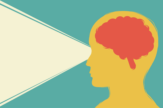
My son is a treasure trove of trivia knowledge. At twelve years old, he is continually scanning the internet for new and unique facts. That’s his hobby and obsession. So, he routinely asks me fun facts about the eyes. One day, he just blurted out, “what part of the brain controls vision?”
I gave him the answer to his question and thought it might be a good topic to share since you might wonder the same thing.
Vision is a complex activity that employs different brain parts to perceive sight, process what you’re seeing, and act on it appropriately. Here’s how it all works…
Which Part of the Brain Controls Vision?
Visual functions are mostly controlled in the occipital lobe of the brain. The occipital lobe is a small area in the brain in the back of the skull. That said, there are many working parts of the brain that combine forces to help you see.
How Your Eyes Work with Your Brain
For instance, when you look at your phone, a particular part of your brain called the pons controls your eye muscles to direct your gaze.
The image of your phone then gets sent through your pupils. It hits the photoreceptor cells in the macula at the back of the eye, including rods (cells responsible for your peripheral vision and night vision) and cones (cells responsible for your color vision and sufficient vision detail).
Rods and cones fire a nerve impulse through the optic nerve, which carries the impulse to a structure at the back of your brain called the occipital lobe.
The occipital lobe processes the image of your phone. However, this is not the end of the sight process.
Next, the visual data is sent to the parietal lobe, which is responsible for giving awareness of the physical distance between you and the phone and your depth perception.
The visual data is also sent to the temporal lobe, which is associated with memory. It recognizes that what you’re looking at is a phone and not a shoe or anything else but a phone.
The temporal lobe is responsible for giving meaning to what we see—it’s what helps you use your phone for its intended purpose, rather than eating it or using it as a shoehorn.
Also, there is evidence that the frontal lobe, which is the reasoning/thinking part of our brain, is involved in the process of vision. The frontal lobe is responsible for maintaining your focus. In this case, it holds your attention on your phone and only your phone and not anything else around it.
So, you may ask yourself, “If the different parts of the brain handle vision, then what about the other aspects of your vision like color or peripheral vision?”
What Part of the Brain Controls Color Vision?
There is a particular part of the occipital lobe that handles color vision. It’s called the visual cortex. There are two visual cortexes, a left and a right one on each occipital lobe.
For example, when you see an apple, the apple’s light gets picked up by the photoreceptors inside the eye (in the retina). Rods and cones receive the wavelengths of light given off by the apple—especially the cones since they’re the eye’s color receptor.
The cones send the impulse through the optic nerve to the visual cortex in the occipital lobe. The brain processes this input based on how many cones were activated and the signal’s strength. That’s how you see the color of the apple is red.
What Part of the Brain Controls Peripheral Vision?
Let’s start with what peripheral vision is… It’s that part of your vision that’s off to the edge of your central gaze. It could be to the left, right, up, or down relative to your central vision.
Our ability to see peripheral vision lies in the fovea, which is at the center of the macula inside the eye. Within the central fovea are rods and cones, with rods making up most of the perifoveal (most peripheral fovea). Rods are more tuned to movement, shapes, and forms rather than fine detail.
This means that when you see something off to the side of your field of vision, you don’t see it as well as if it were right in front of you. It correlates with the composition of the fovea—the center of the fovea contains rods and cones, which means your central vision is more detailed. On the other hand, the peripheral fovea is primarily rods, so your peripheral vision is limited by the function of rods which do not convey sharp detail.
The rods’ impulses in the perifovea travel along the optic nerve to the anterior visual cortex in the occipital lobe for processing.
Final Thoughts
Vision is a very complicated activity for the brain to process. The fact that all the visual information goes through the eye to the optic nerve and then to the brain in a fraction of a second is nothing short of incredible.
We mostly perceive the world through our sight, and so it is a precious gift that we must not take for granted.


